In general, aneuploidy refers to an abnormal number of chromosomes in a cell or organism, which can result in significant developmental and physical changes. Most of these chromosome mis-segregation events lead to the death of the cell or embryo, as the complex relationship between genes and regulatory elements in the genome is disturbed. The following list describes some of the few common types of whole-chromosome aneuploidies seen in babies born.
This is only a general overview, not a scientific write-up or medical advice and no judgement about any condition mentioned.
1. Trisomy 21 (Down syndrome)
The most common chromosomal disorder, occurring in approximately 1 in every 700 to 1,000 live births worldwide, is characterized by the presence of an extra copy of chromosome 21. Children with Down syndrome typically have characteristic facial features, intellectual disability, developmental delays, and an increased risk of certain medical conditions like congenital heart defects and leukemia.
2. Trisomy 18 (Edwards syndrome)
An extra copy of chromosome 18 occurs in about 1 in every 5,000 live births worldwide. Children with Edwards syndrome often have severe developmental delays, intellectual disability, distinctive facial features, heart defects, and other abnormalities of various organs. Infants with trisomy 18 have a high mortality rate, only a small percentage survive beyond their first year of life.
3. Trisomy 13 (Paetau syndrome)
An extra copy of chromosome 13 is estimated to occur in approximately 1 in every 10,000 to 20,000 live births worldwide. Children with Patau syndrome typically have severe intellectual disability, multiple physical abnormalities (cleft lip and palate, heart defects, and brain malformations), and a high mortality rate in infancy.
4. Monosomy X (Turner syndrome)
Monosomy X, also known as Turner syndrome, occurs when a female is born with a single X chromosome instead of the usual two. It affects approximately 1 in every 2,500 to 5,000 live female births. Girls with Turner syndrome often have short stature, delayed puberty, infertility, and may experience a range of other health issues, including heart defects, kidney problems, and learning difficulties.
5. XXY (Klinefelter syndrome)
XXY is characterized by the presence of an extra X chromosome in males and occurs in approximately 1 in every 500 to 1,000 live male births worldwide. Boys and men with Klinefelter syndrome often have tall stature, hypogonadism (reduced testosterone production), infertility, learning difficulties, and may experience other physical and behavioral differences.
6. XYY (Jakobs syndrome)
XYY males show only mild symptoms, including tall stature and an increased risk of learning disabilities. It occurs in about 1 in 1,000 male births.
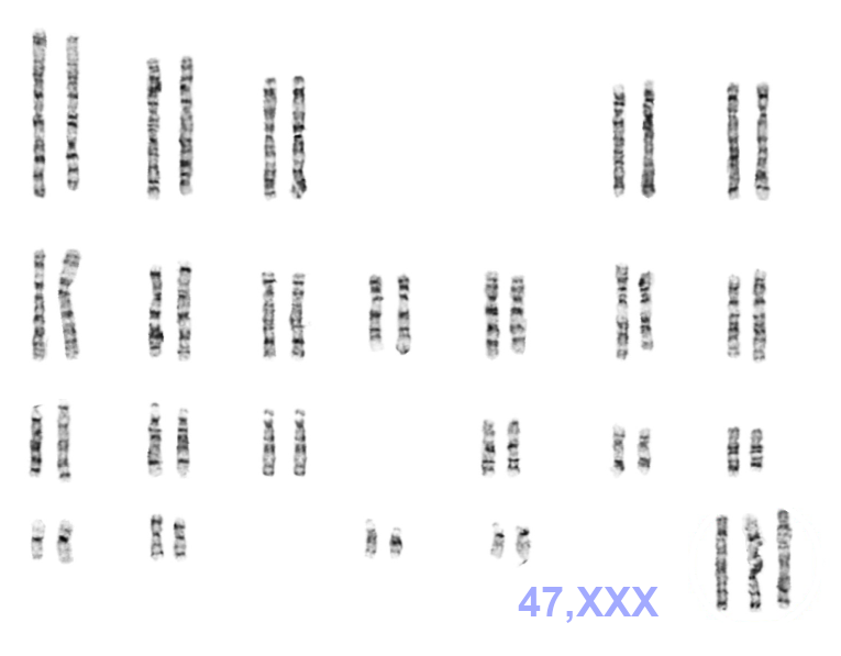
7. Trisomy X (Triple X syndrome)
The effects of XXX on the affected female carrier range from no symptoms to significant disability and often involve learning disabilities, mild dysmorphic features such as hypertelorism (wide-spaced eyes) and clinodactyly (incurved little fingers), early menopause, and increased height. The occurrence is around 1 in 1,000 female births.
Because of limited testing options in some countries and because of the variability of the symptoms, the actual prevalence of these genotypes might be higher.
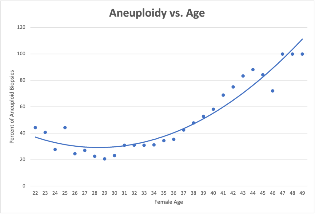
As mentioned, all chromosomes can be affected by these distribution errors during early development, most are not viable at all however. The number of chromosomal segregation errors leading to aneuploidy and fertility problems generally increase with the maternal age (see graph), although these could of course alternatively be linked to inherited chromosomal changes from the mother’s or the father’s side as well as completely different reasons.
Advances in prenatal screening and diagnostic techniques have allowed for early identification of some of these chromosomal abnormalities, leading to better support and care for affected individuals and their families.
The possibilities to detect these chromosomal distribution errors as part of an IVF treatment have improved over the last decade, reducing the number of failed implantation attempts and therefore the costs and agony for patients attempting to conceive.

Graph produced with data from: Franasiak, et al. “The nature of aneuploidy with increasing age of the female partner: a review of 15,169 consecutive trophectoderm biopsies evaluated with comprehensive chromosomal screening”, Fertility and Sterility 2014
Further links for the general public:


